Map of Zika virus reveals how it shifts as it matures
Before an immature Zika virus becomes infectious, it does some major remodeling.
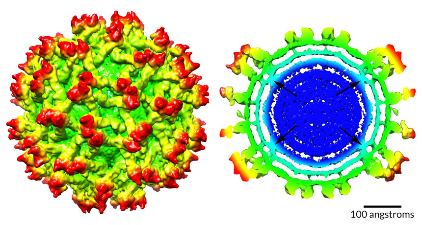
In a fledgling virus particle, the inner protein and RNA core (shown in dark blue above, right) forms bridges to the membrane layer that surrounds it. As the virus matures, the core shuffles around and the bridges melt away (below, right).
It’s the first time scientists have seen such rearrangement in the core of a flavivirus, the group that also includes the viruses that cause dengue, West Nile and yellow fever, says virologist Richard Kuhn of Purdue University in West Lafayette, Ind.
Scientists don’t know why the immature Zika virus reshuffles its insides, Kuhn says — perhaps it helps the maturing virus become infectious. But that’s the next big question to answer, he says.
If blocking the reorganization somehow made mature viruses harmless, scientists would have a new clue about preventing Zika infection. Kuhn and colleagues’ map of the immature virus’s structure, published online January 9 in Nature Structural & Molecular Biology, could offer other hints for thwarting Zika.
With a technique called cryo-electron microscopy, the team could see three-headed protein spikes (shown in red) studding the surface like some kind of medieval weapon, and could even distinguish the separate layers of the membrane (aqua) that encloses the core. (The maps are radially colored; colors change as distance from the core increases.) Outside the membrane lie surface proteins called envelope, or E, proteins (green and yellow) that help the virus sneak into cells.
Last year, Kuhn’s team reported the structure of the mature Zika virus (SN: 4/30/16, p. 10). The new work offers another illuminating peek at Zika — a baby picture, of sorts.